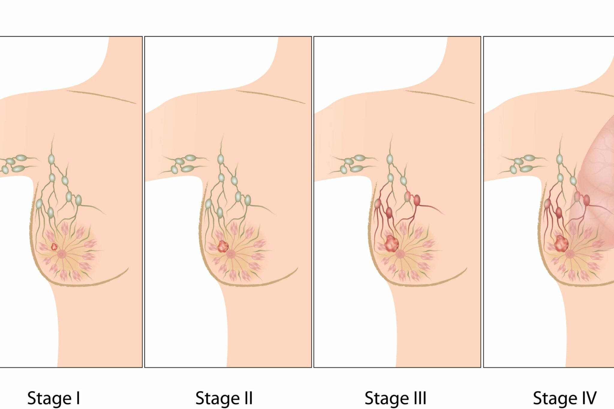
Diagnosing and Treating Breast Cancer During Pregnancy

Despite being the second most common malignancy affecting pregnancy, breast cancer during pregnancy is rare. Known as Pregnancy-Associated Breast Cancer (PABC), it affects approximately one in 3000 pregnancies. PABC is, in fact, defined as breast cancer that is diagnosed during pregnancy or in the first year postpartum.
The aim with diagnosis and treatment of breast cancer during pregnancy is to follow, as closely as possible, the normal standards of care for patients of the same age who are not pregnant. That being said, some modifications may be required in order to minimise risks to the developing baby.
Diagnosis
Most breast cancers are diagnosed following self-examination and identification of a lump in the breast tissue. During pregnancy, the breasts change in shape and size as they prepare for breastfeeding. It is not uncommon for them to feel lumpy and inconsistent as the milk producing ducts and glands start to fill with milk. This can make it difficult for women to establish which changes are normal and which are a cause for concern. All abnormal masses should be investigated, although, fortunately, 80% will be benign in nature.
The first diagnostic test a doctor will use is ultrasound. This uses soundwaves and is entirely safe for the unborn baby. It will characterise any unusual masses and identify whether there are features of concern within the mass. At around the same time a needle aspiration and/or core biopsy may be taken. This enables doctors to explore the cells of the breast in more detail. Particular care will need to be implemented for analysing the results as, during pregnancy, it is not unusual for cells to become more proliferative in nature. Rapidly proliferating cells under normal conditions can serve as a warning sign that something is amiss.
Mammograms will be used, however, they are known to lack sensitivity in pregnant or lactating females. As a mammogram involves radiation, doctors will endeavor to shield the baby from exposure. Newer digital mammograms might improve the sensitivity of the technique in women under 40 years of age.
CT scans and bone scans, which are a normal part of the diagnostic process in non-pregnant females, are avoided during pregnancy, due to the dangers of radiation to the developing foetus. These methods are normally employed to check for metastasis, and thus, metastatic disease can be harder to detect in pregnant women.
Treatment
Most women with PABC will undergo surgery as the first-line treatment option, usually in the form of a modified radical mastectomy. It is generally safe to undergo anaesthesia whilst pregnant, but in order to limit the time you are under general anaesthetic, your doctor will probably recommend postponing reconstructive surgery until after delivery.
Radiotherapy is not recommended during pregnancy and wherever possible your doctor will attempt to delay this type of treatment until after delivery. This is because it increases the risk of foetal malformations and can delay neurocognitive development. If breast preservation is desired, the disease is not advanced and diagnosis has occurred towards the end of pregnancy, it may be possible to treat with immediate lumpectomy and radiotherapy after delivery. It has been shown that a six week window between lumpectomy and commencement of radiotherapy does not have a detrimental effect on outcome.
Chemotherapy should not be given during the first 14 weeks of pregnancy. It can cause severe teratogenicity during organ development, which primarily occurs in the first trimester. In the second and third trimester, chemotherapy can be administered. There have been no reports of later ill effects in children born to mothers who had chemotherapy at this stage of their pregnancy. Most doctors will recommend stopping chemotherapy at about week 36 to reduce the risk of infection or bleeding during delivery.
Hormone therapy is not recommended for women who are pregnant or breastfeeding.
Whilst, there are treatment options for women with PABC, additional work is required to establish a more effective treatment approach for these women. Certainly, as women postpone having children, the rates of PABC are likely to increase over the next few years.
Prognosis
The prognosis for women with PABC is generally lower than for women with breast cancer who are not pregnant. This is likely due to:
- Less aggressive therapy being used due to concerns over the effect of harsher regimens on the developing baby.
- Later stage of diagnosis because of difficulty in distinguishing physiologically-relevant changes from normal pregnancy-related changes.
- The pregnancy having a direct effect on outcome, although knowledge regarding the exact mechanisms relating to this is currently lacking.
- An increased percentage of oestrogen receptor negative cases. This is known to be associated with an increased risk of metastatic disease, which has a poorer prognosis.
In terms of the developing foetus, women with PABC should be reassured that there are no reports of breast cancer spreading from the mother to the baby during pregnancy. In rare cases cancer cells will be found in the placenta, so the doctor will always check this immediately after delivery.
During pregnancy, a woman should remain under observation by a multidisciplinary team of healthcare professionals, including gynaecologists and oncologists to ensure that the support she receives is optimal for both her and her baby. Growth scans will be performed regularly to ensure that the baby is developing as he or she should be. If possible, the medical team will try to ensure that the woman delivers her baby as close to her due delivery date as possible. After delivery, treatment options will be reassessed.
Get yourself the post-surgery pack
Nabta is reshaping women’s healthcare. We support women with their personal health journeys, from everyday wellbeing to the uniquely female experiences of fertility, pregnancy, and menopause.
Get in touch if you have any questions about this article or any aspect of women’s health. We’re here for you.
Sources:
- “Breast Cancer.” Breast Cancer during Pregnancy | Cancer Research UK, 21 Nov. 2017, https://www.cancerresearchuk.org/about-cancer/breast-cancer/living-with/breast-cancer-during-pregnancy.
- “Breast Cancer, Pregnancy and (Green-Top Guideline No. 12).” Royal College of Obstetricians & Gynaecologists, https://www.rcog.org.uk/en/guidelines-research-services/guidelines/gtg12/.
- Johansson, A L V, et al. “Diagnostic Pathways and Management in Women with Pregnancy-Associated Breast Cancer (PABC): No Evidence of Treatment Delays Following a First Healthcare Contact.” Breast Cancer Research and Treatment, vol. 174, no. 2, Apr. 2019, pp. 489–503., doi:10.1007/s10549-018-05083-x.
- Keyser, E A, et al. “Pregnancy-Associated Breast Cancer.” Reviews in Obstetrics & Gynecology, vol. 5, no. 2, 2012, pp. 94–99.


