What is a Hysteroscopy?


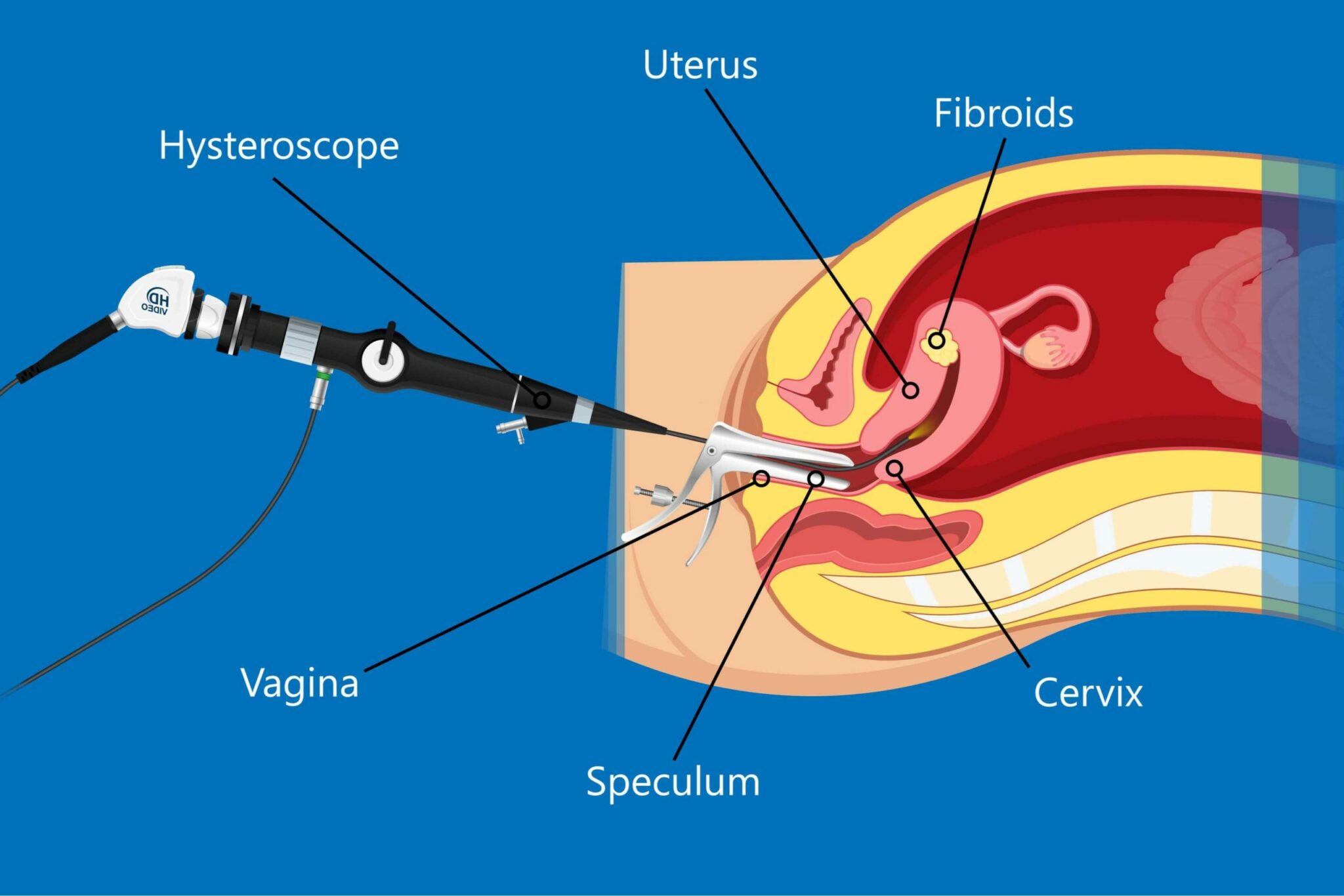
Hysteroscopy is a means of examining the inside of the uterus, without having to make any cuts to the abdomen. A narrow, lighted tube with a camera attachment is inserted via the vagina and cervix and used to assess the contents and lining of the uterus. The process can be diagnostic or operative and is described as being ‘accurate, rapid, gentle and safe’. Many women report only mild discomfort and fewer than 1% have post-procedure complications.
The procedure involves inserting the hysteroscope and then filling the uterus with carbon dioxide gas, or liquid to expand it and clear it of blood and mucus. This gives the doctor an unimpeded view of the inside of the uterus.
Diagnostic Hysteroscopy
Hysteroscopy can be used to evaluate unexplained bleeding, heavy periods or post-menopausal bleeding. For women struggling with unexplained infertility, it can be used to identify uterine pathology that is preventing pregnancy or causing repeated miscarriages. It can be used to diagnose non-cancerous growths within the uterus, including polyps and fibroids. Modern, high resolution hysteroscopes can also be used to diagnose endometriosis, which can cause chronic pain and infertility.
The advantage of using a hysteroscope for diagnostic, evaluation or exploratory purposes is that anaesthetic is seldom required and the procedure can be completed in under 20 minutes. Cramping and spotting are common, but more severe side effects are very rare.
Operative Hysteroscopy
Sometimes a doctor will attempt to treat a condition at the same time as diagnosing it to avoid the need for additional procedures. This happens regularly when fibroids or polyps are identified in the uterus. The doctor will pass fine surgical instruments along the hysteroscope, with the aim of excising the unwanted growths. Hysteroscopy can also be used to remove uterine adhesions, which are bands of scar tissue that can provide a structural barrier to conception.
If the doctor is using a hysteroscope to examine the endometrium, he or she my choose to take a biopsy at the same time, so that the cells of the uterus lining can be examined in more detail. This avoids the need to carry out a more invasive procedure, such as a laparoscopy, which involves making an incision into the wall of the abdomen.
An hysteroscopy can be performed in place of a dilation and curettage (D & C) to locate and remove any products that are retained in the uterus following a miscarriage.
It can also be used to destroy the uterine lining, via endometrial ablation, as a treatment option for women experiencing heavy bleeding.
In conclusion, hysteroscopy is a valuable tool in women’s health care. It can be used to identify and treat existing structural problems and is minimally invasive in nature.
Nabta is reshaping women’s healthcare. We support women with their personal health journeys, from everyday wellbeing to the uniquely female experiences of fertility, pregnancy, and menopause.
Get in touch if you have any questions about this article or any aspect of women’s health. We’re here for you.
Sources:
- “Hysteroscopy.” NHS Choices, NHS, www.nhs.uk/conditions/hysteroscopy/.
- “Hysteroscopy.” Cleveland Clinic, my.clevelandclinic.org/health/treatments/10142-hysteroscopy.
- Parry, J. Preston, and Keith B. Isaacson. “Hysteroscopy and Why Macroscopic Uterine Factors Matter for Fertility.” Fertility and Sterility, vol. 112, no. 2, 2019, pp. 203–210., doi:10.1016/j.fertnstert.2019.06.031.


